Syphillis
by [The Seventh Nazgûl ]
DISCLAIMER: I am still an undergraduate medical student who is not yet fully qualified to practice medicine, as I have still much to learn. For the articles I write here, I use some of the most reliable textbook and online sources. Thus I am only advising people, not treating them whatsoever. If you have read anything in my articles, don't go ahead and use it in accordance, for though the information is reliable, the need to see a fully qualified legally certified medical practitioner is mandatory. Also, note that medicine is an ever-advancing science, for example what might have been medically acceptable this month may become an out-of-date practice next week if a new and improved regimen takes its place. This said, these articles are only written for the sole purposes of information, myth-busting, and advice. They do not override the necessity of seeing a doctor if your health is at stake.
WARNING: At the end of this article there are some clinically intimate pictures which show the affected genitalia and other body parts of patients. They are meant for informative and scientific purposes. Steel yourself if you cannot stand the sight of organs damaged by diseases or if you are offended by seeing genitalia.
Introduction
Syphilis is one of the most ancient sexually transmitted diseases (STDs), known since the renaissance and medieval times. Syphilis is a chronic (lasting for many years) slowly progressive worldwide disease, the third commonest STD and is primarily transmitted by sexual contact. Transmission from pregnant females to their foeti has been observed.
Causative Agent
The causative organism of syphilis is a long, thin, spiral, and motile (i.e. it can move using special structures) Gram negative bacterium called Treponema pallidum (figure 1). It is a fragile organism that can be killed by soap and water or heat (before the discovery of antibiotics, syphilis used to be treated by fever boxes, a metallic box in which the patient was placed in sunlight with nothing but his/her head showing. The heat from the sun was trapped in the fever box, resulting in accumulation of heat which was supposed to kill the bacteria. This practice is an uncomfortable one and is no longer applicable nor acceptable nowadays). Also, it was discovered that T. pallidum can be killed my mercury, hence the popular medieval saying: An eternity with Venus is worth one night with Mercury.
Clinical Picture
Syphilis is an infectious disease of three stages designated primary, secondary and tertiary.
Primary syphilis occurs after the infection with T. pallidum . At this stage, the organism is present in amounts sufficient to be viewed by special microscopic techniques to confirm the diagnosis.
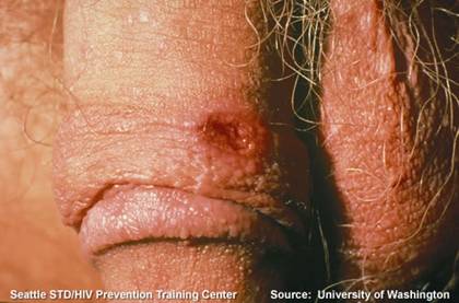
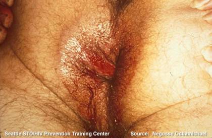
The lesion (damage to the body) characteristic of primary syphilis is called the chancre. It is a painless inflammatory (and highly infectious) ulceration of the skin of the site that came in contact with the bacteria. The chancre appears after a period of a week up to 2 months following infection, with an average period of three weeks.
In most cases, the chancre appears on or around the genitalia (figures 2 and 3), but it appear on other sites such as the mouth [attributed to oral sex (?)] or the hands of healthcare workers who approach a patient's chancre without protective measures and washing hands carefully after handling every patient. The chancre heals spontaneously without scarring and after that the patient enters the secondary stage.
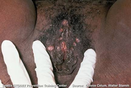
In the secondary stage, the patient is still highly infectious, but the diagnosis is mostly based on the detection of substances specific to the infection in the blood. The patient experiences a rash which spreads all over the body, as well as palms and soles (figure 4), this stage is also characterized by the presence of numerous pale moist swellings called condyloma lata in the armpits and the skin folds between the thighs and genitalia (figure 5). The mouth can be affected and this is demonstrated by mucous patches (figure 6). Again, these lesions heal and approximately 40% of cases progress to tertiary syphilis.
In this stage, the patient suffers from lesions called gummas in the skin, bones, and liver. It is should be remarked that the gumma never heals to its original state. Damage to the central nervous system (brain and spinal cord) appears mostly as dementia, paralysis and walking abnormalities which damage the knees. The heart and blood vessels are affected and this manifests itself with many problems. The patient may die due to cerebral (brain) or cardiovascular (heart and vessels) causes.
It is worth noting that some patients may remain apparently healthy throughout the primary and/or secondary stages, but they start to show the signs of the disease in the tertiary stage.
Treatment and Prevention
With the discovery of antibiotics, it was found out that T. pallidum can be killed by a form of penicllin called penicillin G. It is highly curative in the primary and secondary stages of the disease.
There is vaccine against syphilis and prevention depends on responsible and safe sexual intercourse as well as careful examination and handling of patients if you're a healthcare worker (especially a physician or nurse), not to mention proper hand washing (preferably with disinfectant or antiseptic soap) after dealing with each patient.
That said, always make sure that the ground for sexual intercourse is clear and that the practice is safe. One can never be careful enough around this area and a period of pleasure could lead to a lifetime of pain if one practices irresponsible sex.
Next time, an infectious agent more popular with the ladies is our feature, Trichomonas vaginalis .
Figures:

Figure 1, Treponema pallidum as observed by darkfield microscopy. Credit for this image goes to the University of Washington and the Seattle STD/HIV Prevention Training Center.

Figure 2, note the chancre on the penis. Credit for this image goes to the University of Washington and the Seattle STD/HIV Prevention Training Center.

Figure 3, note the chancre at the circumference of the anus. Credit for this image goes to Negusse Ocbamichael and the Seattle STD/HIV Prevention Training Center.

Figure 4, Rash of secondary syphilis. Credit for this image goes to Connie Celum, Walter Stamm, and the Seattle STD/HIV Prevention Training Center.

Figure 5, note the condyloma lata around the external genitalia in secondary syphilis. Credit for this image goes to Connie Celum, Walter Stamm, and the Seattle STD/HIV Prevention Training Center.

Figure 6, secondary syphilis; note the mucous patches on the patient's tongue. Credit for this image goes to the University of Washington and the Seattle STD/HIV Prevention Training Center.
References:
Lippincott's Illustrated Reviews: Microbiology
http://depts.washington.edu/nnptc/online_training/std_handbook/gallery/index.html
Have you read the DISCLAIMER at the top of the page?